


TATORT HAUT – Unter Verdacht BPDCN
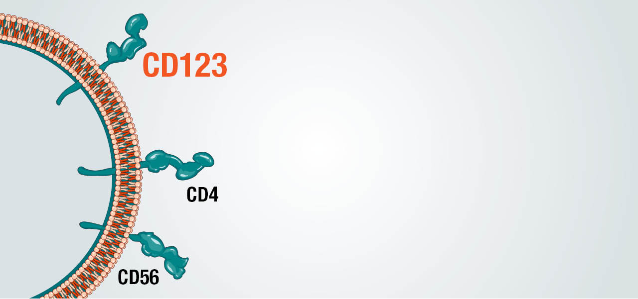
BPDCN ist eine seltene, aggressive Krebserkrankung und erst seit 2016 unter diesem Namen bekannt.1,2 Trotz charakteristischer Marker ist die Diagnose dieser seltenen Erkrankung oft wie eine Detektivarbeit. Aufgrund des heterogenen Erscheinungsbilds der BPDCN in der Klinik3–5 führt erst ihr pathologischer Fingerabdruck zur Lösung und damit zur gesicherten Diagnose.
Deshalb ist für die Ermittlung der Diagnose genaues Hinschauen und Hinterfragen so wichtig – es braucht Krankheitsversteher! Und die Aufmerksamkeit aller!
ZEIT IST DER ENTSCHEIDENDE FAKTOR
Die Seltenheit der BPDCN und Ähnlichkeit zu anderen hämatologischen und dermatologischen Neoplasien stellt eine große Herausforderung für die Diagnose der BPDCN dar.3,4,6,7
Die Verzögerung der Diagnose kann einen negativen Einfluss auf die Prognose haben, da die BPDCN ohnehin eine klinisch sehr aggressive Erkrankung ist.2,6,8,9
Der Austausch zwischen Hausärzten, Dermatologen, Laborarbeitern, Pathologen, Referenzpathologien und Hämato-Onkologen spielt eine wesentliche Rolle bei der korrekten und rechtzeitigen Diagnose von BPDCN.
DAS INDIZIEN-BOARD
DER PATIENT STELLT SICH VOR

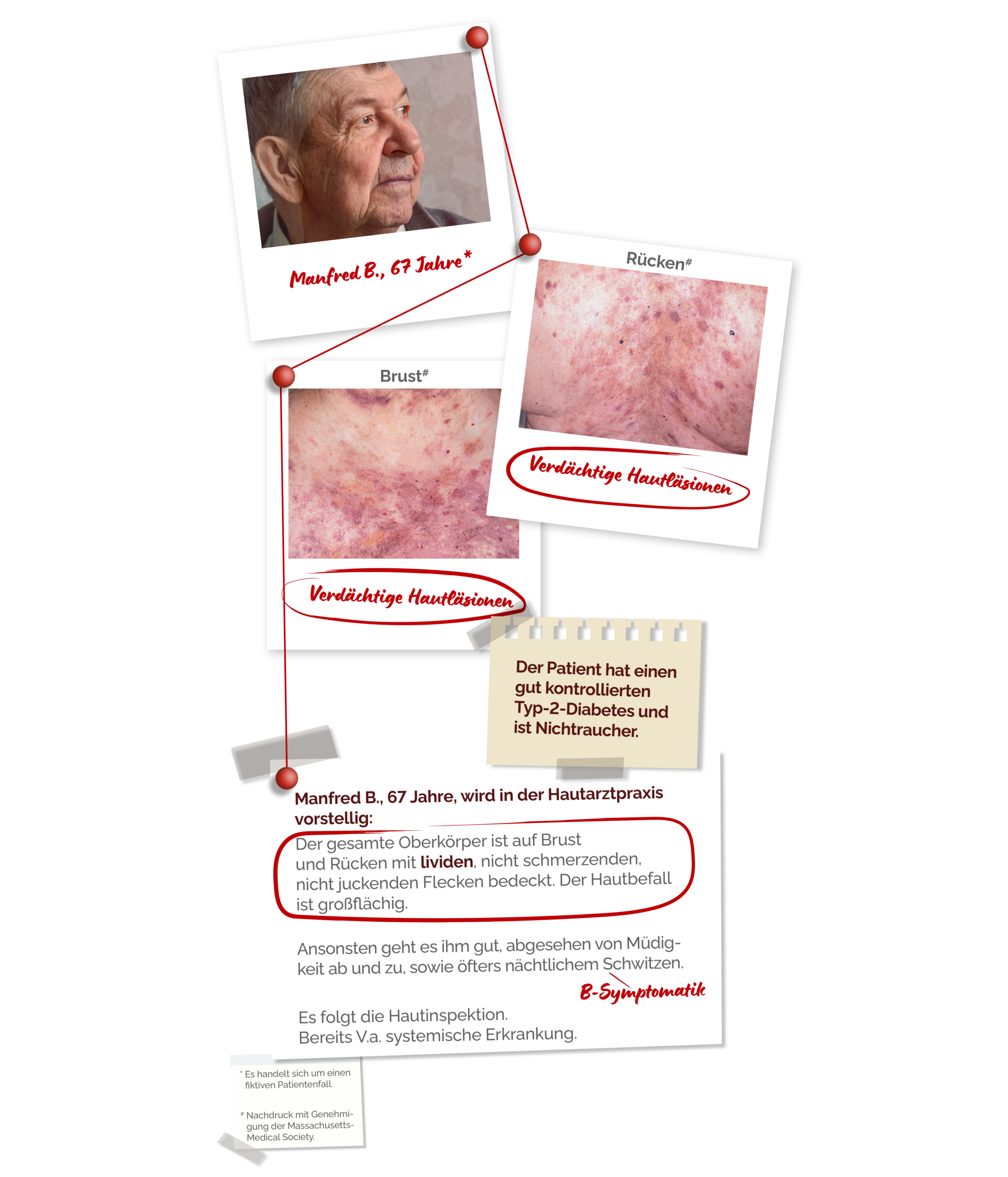
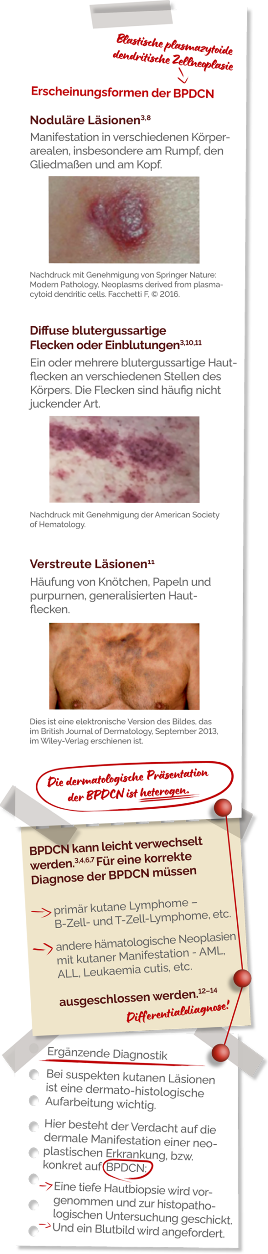
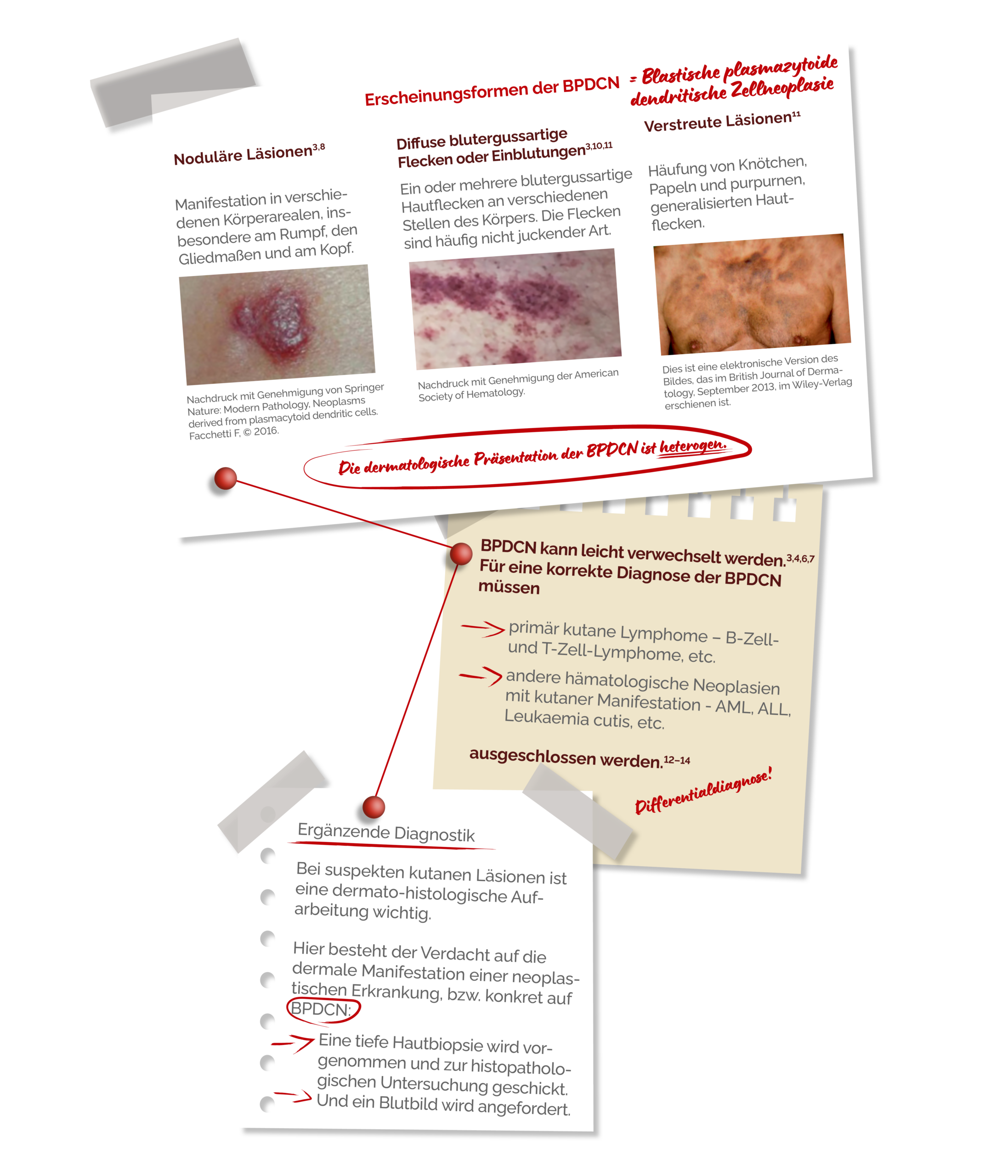
FRÜHERKENNUNG UND BLICKDIAGNOSE
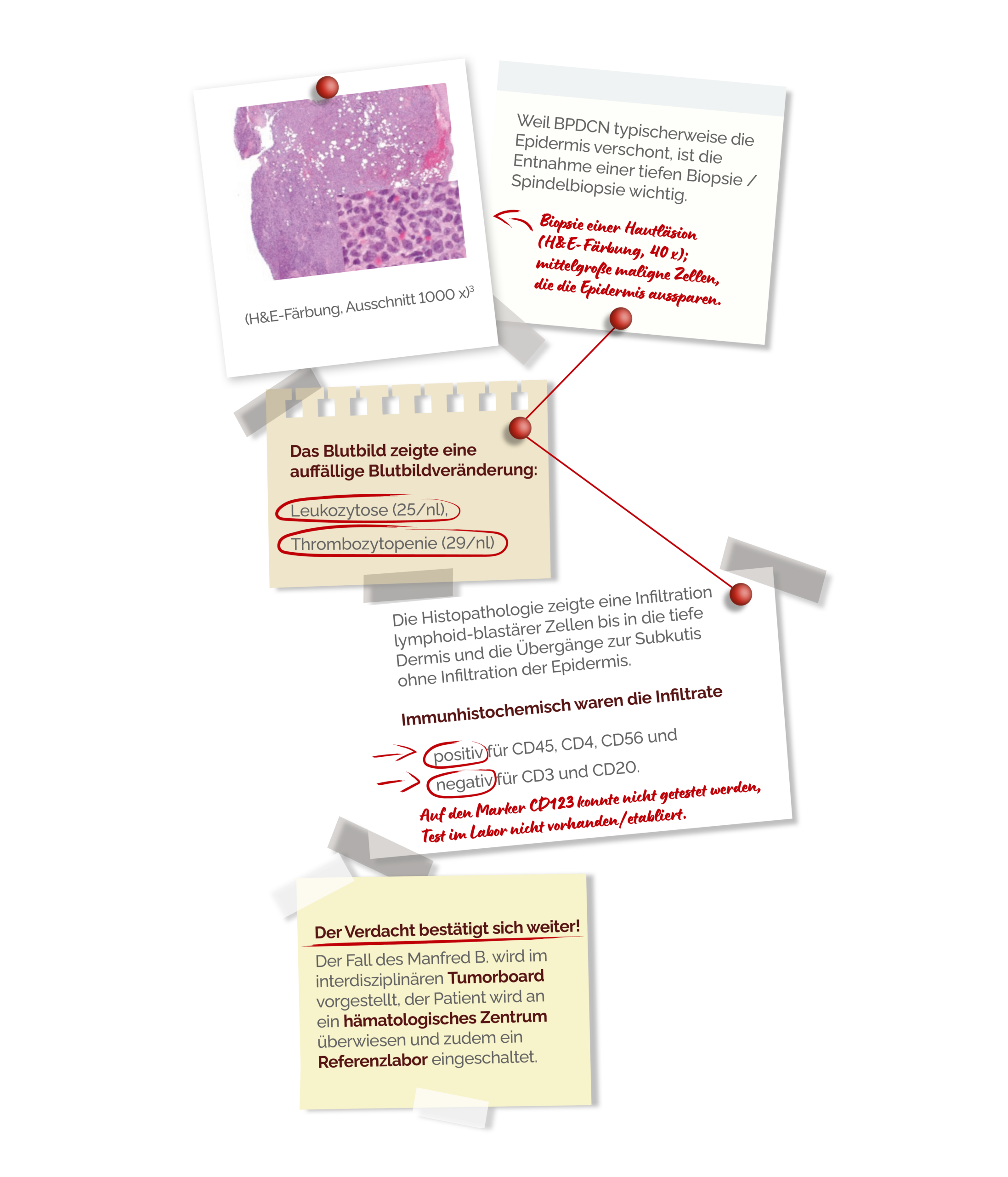
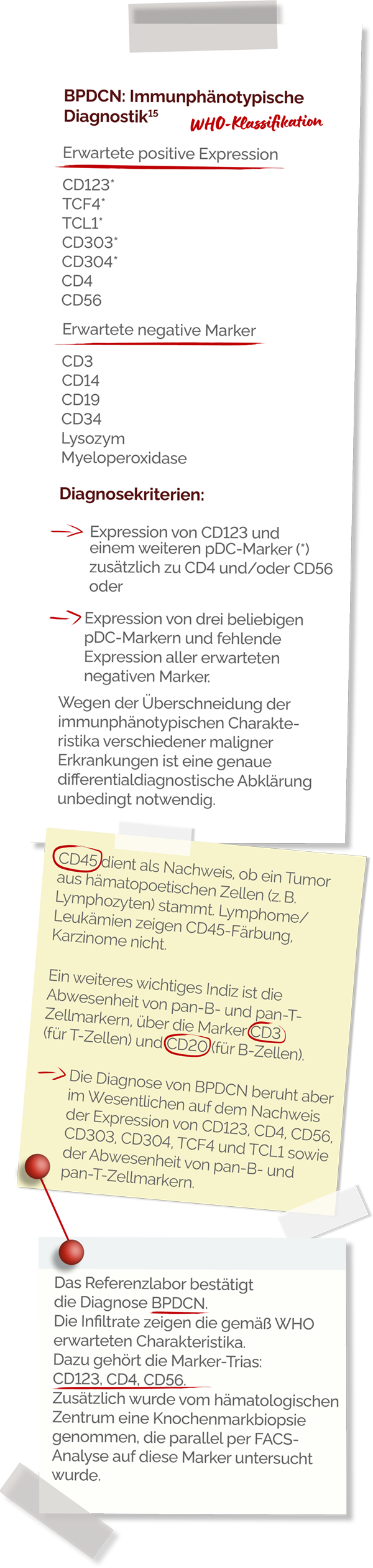
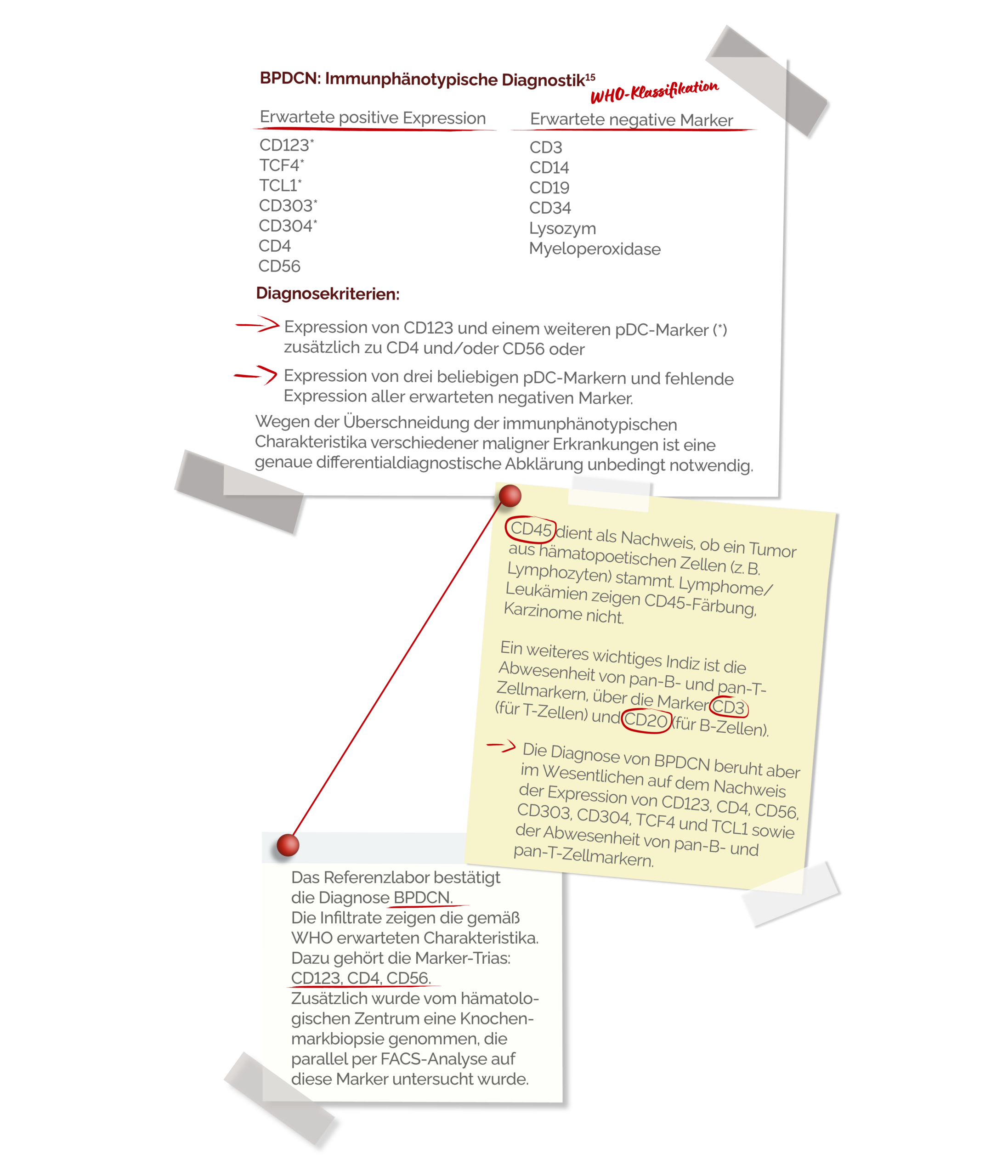
HISTOPATHOLOGIE & ERSTES BLUTBILD

REFERENZLABOR UND DIAGNOSE
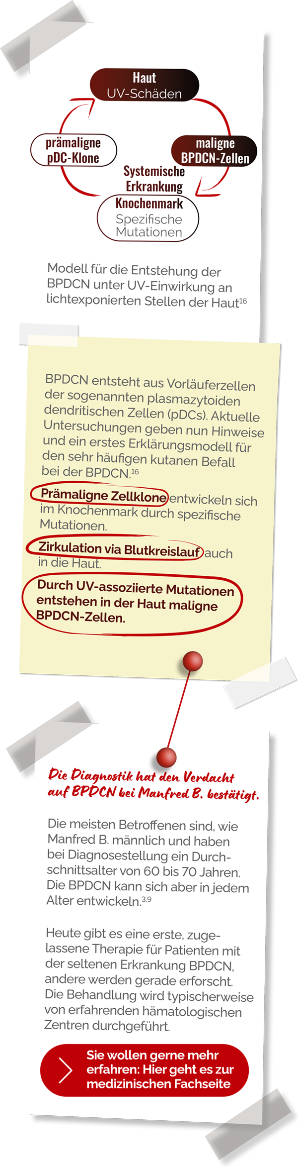
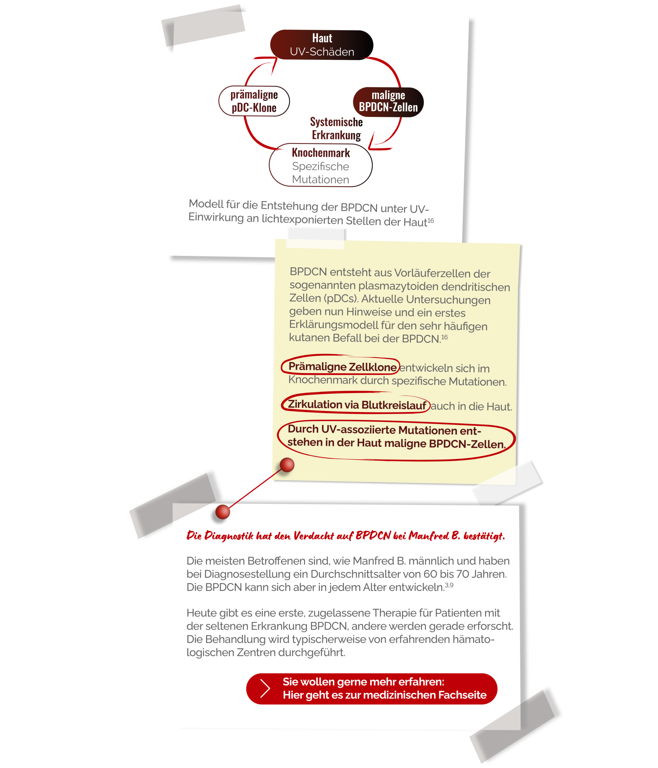
WAS KÖNNTE PASSIERT SEIN?